

Introduction
Mycology is the scientific study of fungi, encompassing yeasts, molds, mushrooms, and other related organisms that play essential roles in nature, medicine, food, and disease prevention. This comprehensive mycology guide for beginners explores the basics of mycology, its significance in microbiology and medicine, major types of fungi, laboratory techniques, common infections, and prevention strategies.
What Is Mycology? (Simple Definition)
Mycology is a branch of microbiology that focuses on the biology, classification, genetics, ecology, and medical relevance of fungi.
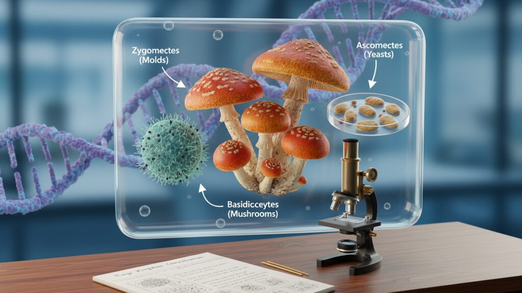
Fungi are distinct organisms that belong to their own kingdom—they are neither plants nor animals. The main groups include:
- Yeasts: Single-celled fungi, e.g., Candida spp.
- Molds: Filamentous fungi, e.g., Aspergillus spp.
- Mushrooms: Macroscopic fruiting bodies
- Dimorphic fungi: Can switch between yeast and mold forms, e.g., Histoplasma spp.
Mycologists study how fungi:
- Grow and reproduce
- Cause disease
- Contribute to medicine, food, and biotechnology
Why Is Mycology Important in Medicine and Microbiology?
Fungi are medically significant because they can cause infections ranging from mild skin issues to life-threatening systemic diseases.
Key Reasons Mycology Matters:
- Rising incidence of invasive fungal infections in immunocompromised patients (HIV, transplant recipients, ICU patients)
- Antifungal resistance, e.g., Candida auris, is a global health threat
- Source of life-saving antibiotics, e.g., penicillin from Penicillium
- Yeasts like Saccharomyces cerevisiae are used in baking and brewing
- Essential for ecosystem nutrient recycling through decomposition
Major Types of Fungi in Mycology
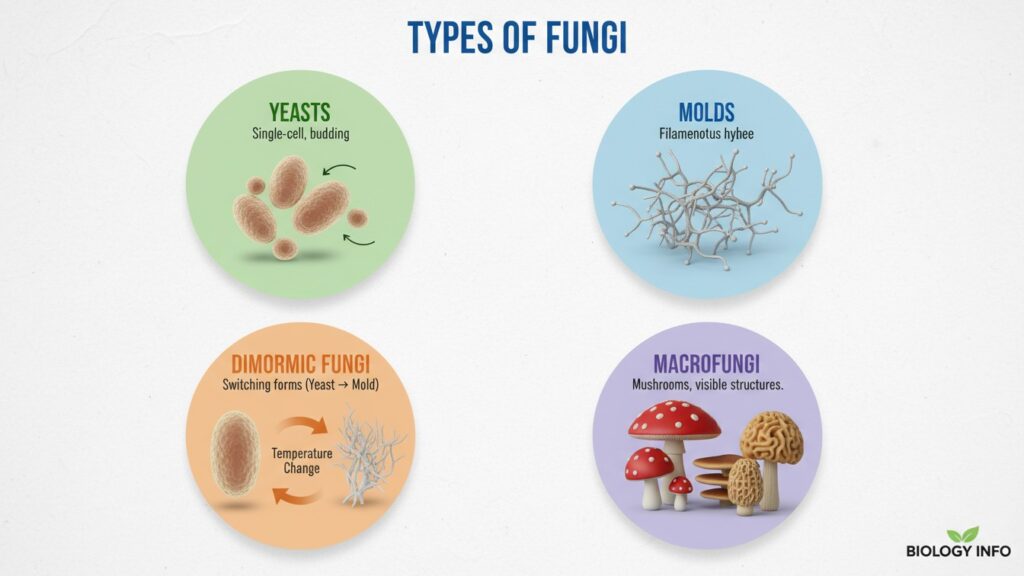
Fungi are classified based on structure, reproduction, and clinical importance.
| Fungal Type | Characteristics | Examples & Importance |
|---|---|---|
| Yeasts | Single-celled, oval/round, reproduce by budding | Candida albicans (oral thrush, vaginal candidiasis), Cryptococcus neoformans (meningitis), Malassezia spp. (skin infections) |
| Molds | Filamentous, grow as hyphae forming mycelium | Aspergillus fumigatus (aspergillosis), Mucor/Rhizopus (mucormycosis), Trichophyton (ringworm) |
| Dimorphic Fungi | Switch forms based on temperature | Histoplasma capsulatum (histoplasmosis), Blastomyces dermatitidis (blastomycosis), Coccidioides immitis (Valley fever) |
| Macrofungi (Mushrooms) | Large visible fruiting bodies | Edible: Agaricus bisporus; Poisonous: Amanita phalloides; Hallucinogenic: Psilocybe spp. |
How Are Fungi Studied in the Lab?

In clinical microbiology, fungi are identified using microscopy, culture, and molecular techniques.
Step-by-Step Lab Techniques
- Sample Collection
- Skin scrapings, nail clippings, hair, sputum, BAL, CSF, blood, tissue biopsies
- Use sterile containers with transport media (e.g., Sabouraud’s transport medium)
- Direct Microscopy
- KOH mount: Dissolves keratin for fungal visualization
- Calcofluor white stain: Binds chitin, fluoresces under UV light
- Gram stain: Yeasts appear Gram-positive
- Culture on Mycological Media
- Sabouraud’s dextrose agar (SDA) – general fungal growth
- Inhibitory mold agar (IMA) – suppresses bacterial growth
- Blood agar – for dimorphic fungi and some yeasts
- Incubation: 25–30°C for molds, 37°C for yeasts/dimorphic fungi
- Macroscopic Examination
- Color: White, green, black, brown, red
- Texture: Powdery, cottony, velvety
- Growth rate: Fast vs. slow growers
- Microscopic Examination
- Lactophenol Cotton Blue mount to observe:
- Hyphae (septate vs non-septate)
- Conidia (spores) arrangement
- Yeast cells and budding patterns
- Lactophenol Cotton Blue mount to observe:
- Biochemical & Molecular Tests
- Germ tube test (Candida albicans)
- Urease test (Cryptococcus)
- MALDI‑TOF MS and PCR for rapid identification
Common Fungal Infections
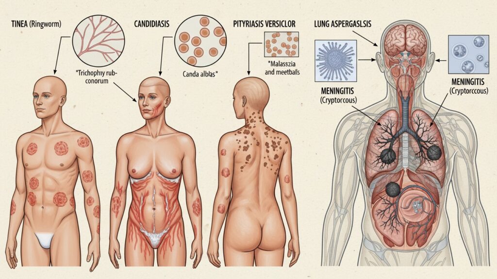
Understanding fungal diseases is critical for diagnosis and treatment.
1. Superficial Fungal Infections
- Tinea (Ringworm): Caused by dermatophytes (Trichophyton, Microsporum, Epidermophyton)
- Types: Tinea capitis (scalp), Tinea corporis (body), Tinea pedis (athlete’s foot)
- Candidiasis: Candida spp. cause oral thrush, vaginal, and cutaneous infections
- Pityriasis Versicolor: Malassezia spp., presents as hypo/hyperpigmented patches
2. Subcutaneous and Systemic Infections
- Aspergillosis: Aspergillus spp. cause ABPA, aspergilloma, invasive infections
- Mucormycosis (Zygomycosis): Mucor/Rhizopus, affects diabetics & immunocompromised, targets sinuses, lungs, brain
- Cryptococcosis: Cryptococcus neoformans, meningitis in HIV patients
- Dimorphic Fungi Infections: Histoplasmosis, Blastomycosis, Coccidioidomycosis, cause pulmonary/systemic infections
Prevention and Control of Fungal Infections
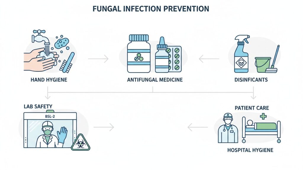
In the Laboratory
- Follow BSL‑2 safety practices
- Wear gloves, a lab coat, and a mask
- Decontaminate surfaces with 70% alcohol or 10% bleach
In Clinical Practice
- Control risk factors: Diabetes, immunosuppression, broad-spectrum antibiotics, and indwelling devices
- Use antifungal prophylaxis for high-risk patients (transplant, neutropenia)
- Educate patients about hygiene for skin/nail infections
Common Mycology Myths vs. Facts
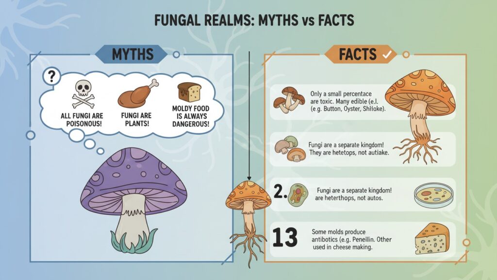
| Myth | Fact |
|---|---|
| “Fungi are just like bacteria.” | Fungi are eukaryotic with nuclei; bacteria are prokaryotic. |
| “All fungi are harmful.” | Many fungi are beneficial (antibiotics, food, decomposition). |
| “Antibiotics work on fungi.” | Antibiotics target bacteria; antifungals are needed for fungi. |
| “Fungal infections are rare.” | Superficial infections are common; invasive infections are rising. |
| “Mycology is only about mushrooms.” | Mycology includes yeasts, molds, and medically important fungi. |
Frequently Asked Questions (Mycology Guide for Beginners)

1. What is the difference between mycology and bacteriology?
Mycology studies fungi (eukaryotes), bacteriology studies bacteria (prokaryotes). Fungi have nuclei, mitochondria, and chitin in cell walls, whereas bacteria lack a true nucleus and have peptidoglycan walls.
2. What are the main tools used in mycology?
- Microscope (KOH, LPCB, Gram stain)
- Sabouraud’s dextrose agar & other fungal media
- Incubators (25–30°C and 37°C)
- Biochemical tests (germ tube, urease)
- MALDI‑TOF MS & PCR for rapid identification
3. How long does it take to identify a fungus in the lab?
- Yeasts: 24–72 hours
- Molds: 5–14 days (slow growers may take weeks)
- Molecular methods: hours to 1–2 days
4. What are the most dangerous fungi in hospitals?
- Candida auris (multidrug-resistant)
- Aspergillus fumigatus (invasive aspergillosis)
- Mucor/Rhizopus (mucormycosis)
- Cryptococcus neoformans (meningitis)
5. Can I specialize in mycology as a microbiologist?
- Yes. Specialists work in clinical labs, research, public health, or pharmaceutical companies, focusing on antifungal drugs and diagnostics.
Conclusion
Mycology, the study of fungi, is vital for medicine, microbiology, food, and ecosystems. Understanding fungi helps prevent infections, combat antifungal resistance, and harness their benefits in biotechnology and daily life. Studying fungi provides essential knowledge for students, researchers, and healthcare professionals alike.
Medical Disclaimer
This content is for informational and educational purposes only and is not a substitute for professional medical advice, diagnosis, or treatment. Always consult a qualified healthcare provider for medical concerns or before starting any treatment.
